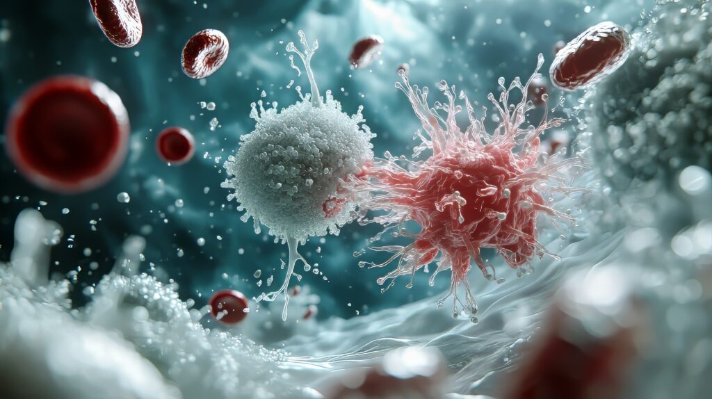
Researchers have identified a mechanism linking inflammation, disease progression, and bone destruction in TCF3::HLF-positive B-cell acute lymphoblastic leukaemia (B-ALL), one of the most aggressive blood cancers affecting children. The findings, published in the Blood Journal, could lead to new therapeutic approaches for this difficult-to-treat malignancy.
A team led by Professor Tomokatsu Ikawa at Tokyo University of Science developed a mouse model that closely mirrors the human disease. Using this model, researchers discovered that leukaemia cells abnormally produce high levels of interleukin-1 beta (IL-1β), an inflammatory protein typically involved in immune responses.
The study found that the TCF3::HLF fusion protein, a genetic abnormality that defines this leukaemia subtype, acts as an abnormal transcription factor, directly activating IL-1β production through a previously unknown regulatory region in the genome. This creates a self-reinforcing loop: IL-1β promotes leukaemia cell proliferation whilst simultaneously activating osteoclasts, the cells responsible for bone breakdown.
When researchers blocked IL-1β using antibodies in diseased mice, leukaemia growth slowed, bone damage decreased, and survival improved. Combining IL-1β inhibition with existing leukaemia treatments produced even stronger effects.
The researchers noted that IL-1β is also actively expressed in other blood cancers, including acute myeloid leukaemia, myelodysplastic syndrome, and multiple myeloma, suggesting broader therapeutic applications.
TCF3::HLF-positive B-ALL is characterised by rapid disease progression, severe bone pain, frequent relapse, and low survival rates. The lack of accurate animal models had previously hindered research progress. The team anticipates early-phase clinical evaluation of IL-1β-targeting treatments within the next few years.
Source: Medical Xpress / Tokyo University of Science (Blood Journal, 2026)